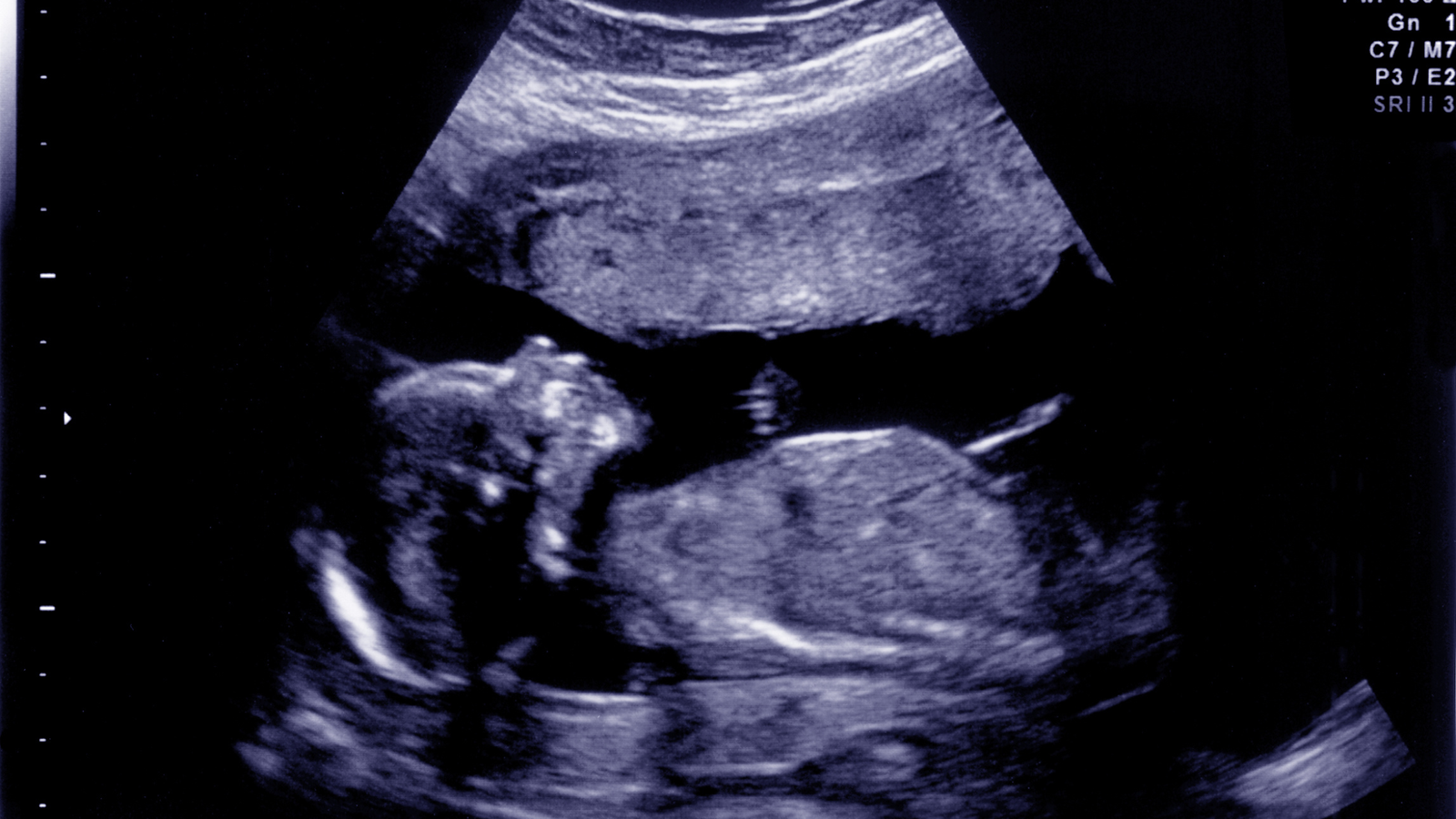
When expecting a baby, every parent wishes for their child to be born healthy and strong. While advancements in medical technology have significantly reduced risks during pregnancy, certain conditions may require specialized attention even before a baby is born. A fetal echocardiogram is one such advanced diagnostic tool that enables doctors to examine a baby’s heart during pregnancy, ensuring any potential concerns are identified and addressed early.
Dr. Nidhi Rawal, a leading pediatric cardiologist in Gurgaon with over 15 years of experience, emphasizes the significance of fetal echocardiograms in safeguarding the health of unborn children. Let’s explore what conditions can be diagnosed with this specialized test and why it is a vital part of prenatal care.
What Is a Fetal Echocardiogram?
A fetal echocardiogram is a specialized ultrasound that evaluates the structure and function of a fetus’s heart. This non-invasive procedure uses sound waves to create detailed images, allowing doctors to study the heart’s anatomy, rhythm, and blood flow patterns. Typically performed between the 18th and 24th weeks of pregnancy, it provides valuable insights into the baby’s cardiovascular health.
When Is a Fetal Echocardiogram Recommended?
A fetal echocardiogram may be recommended in the following scenarios:
- Family History: If there is a history of congenital heart defects in the family.
- Abnormal Ultrasound Findings: When a routine prenatal ultrasound shows potential heart abnormalities.
- Maternal Health Conditions: If the mother has conditions such as diabetes, lupus, or certain infections during pregnancy.
- Use of Certain Medications: If the mother has taken medications linked to an increased risk of congenital heart defects.
- Chromosomal Abnormalities: When genetic tests reveal abnormalities like Down syndrome.
- In-Vitro Fertilization (IVF): Pregnancies achieved through IVF may undergo closer monitoring.
Conditions Diagnosed with a Fetal Echocardiogram
Fetal echocardiography is a powerful tool to detect a wide range of heart conditions in unborn babies. Some of the most common diagnoses include:
1. Congenital Heart Defects (CHDs)
These are structural abnormalities present at birth. Common CHDs diagnosed include:
- Atrial Septal Defect (ASD): A hole in the wall between the heart’s upper chambers.
- Ventricular Septal Defect (VSD): A hole in the wall between the lower chambers.
- Tetralogy of Fallot (TOF): A combination of four heart defects that affect normal blood flow.
- Hypoplastic Left Heart Syndrome (HLHS): Underdevelopment of the left side of the heart.
2. Arrhythmias
Fetal echocardiography can identify abnormal heart rhythms, such as:
- Tachycardia: Abnormally fast heart rate.
- Bradycardia: Abnormally slow heart rate.
- Irregular Heartbeats: Variations in rhythm that may require monitoring.
3. Valve Abnormalities
Issues with the heart valves, such as narrowing (stenosis) or improper closing, can be detected.
4. Heart Tumors
Rarely, benign tumors like rhabdomyomas can be identified during fetal echocardiography.
5. Pericardial Effusion
This condition involves the accumulation of fluid around the heart, which may indicate infection or other complications.
6. Major Vessel Abnormalities
Fetal echocardiography can detect issues with the major blood vessels connected to the heart, such as:
- Transposition of the Great Arteries (TGA): The two main arteries are reversed.
- Coarctation of the Aorta: Narrowing of the Aorta.
Importance of Early Diagnosis
Early detection of heart abnormalities offers several benefits, including:
- Prenatal Planning: Parents and doctors can prepare for specialized care immediately after birth.
- Improved Outcomes: Timely interventions can significantly improve survival rates and quality of life.
- Peace of Mind: Knowing the baby’s condition helps reduce anxiety and uncertainty during pregnancy.
What to Expect During the Procedure
A fetal echocardiogram is a safe and painless procedure that typically takes 30 to 60 minutes. During the test:
- The expectant mother lies down while a specialized technician applies a gel to the abdomen.
- A transducer (probe) is moved over the abdomen to capture images of the baby’s heart.
- The results are reviewed by a pediatric cardiologist, who provides a detailed report and recommendations.
Trust Child Heart Health for Expert Care
At Child Heart Health, Dr. Nidhi Rawal and her team are dedicated to ensuring the best possible outcomes for children with heart conditions. With state-of-the-art technology and compassionate care, Dr. Rawal has earned a reputation as one of the best pediatric cardiologists in Gurgaon. If you’re expecting and have been advised to undergo a fetal echocardiogram, rest assured that your baby’s heart health is in expert hands.
Conclusion
A fetal echocardiogram is a critical tool in modern prenatal care, enabling the early detection and management of heart conditions in unborn babies. Identifying potential issues before birth, it empowers parents and doctors to make informed decisions, ensuring the best possible start for the child. If you have concerns about your baby’s heart health, consult Dr. Nidhi Rawal at Child Heart Health today.
Book your consultation now to secure your baby’s healthy future!


