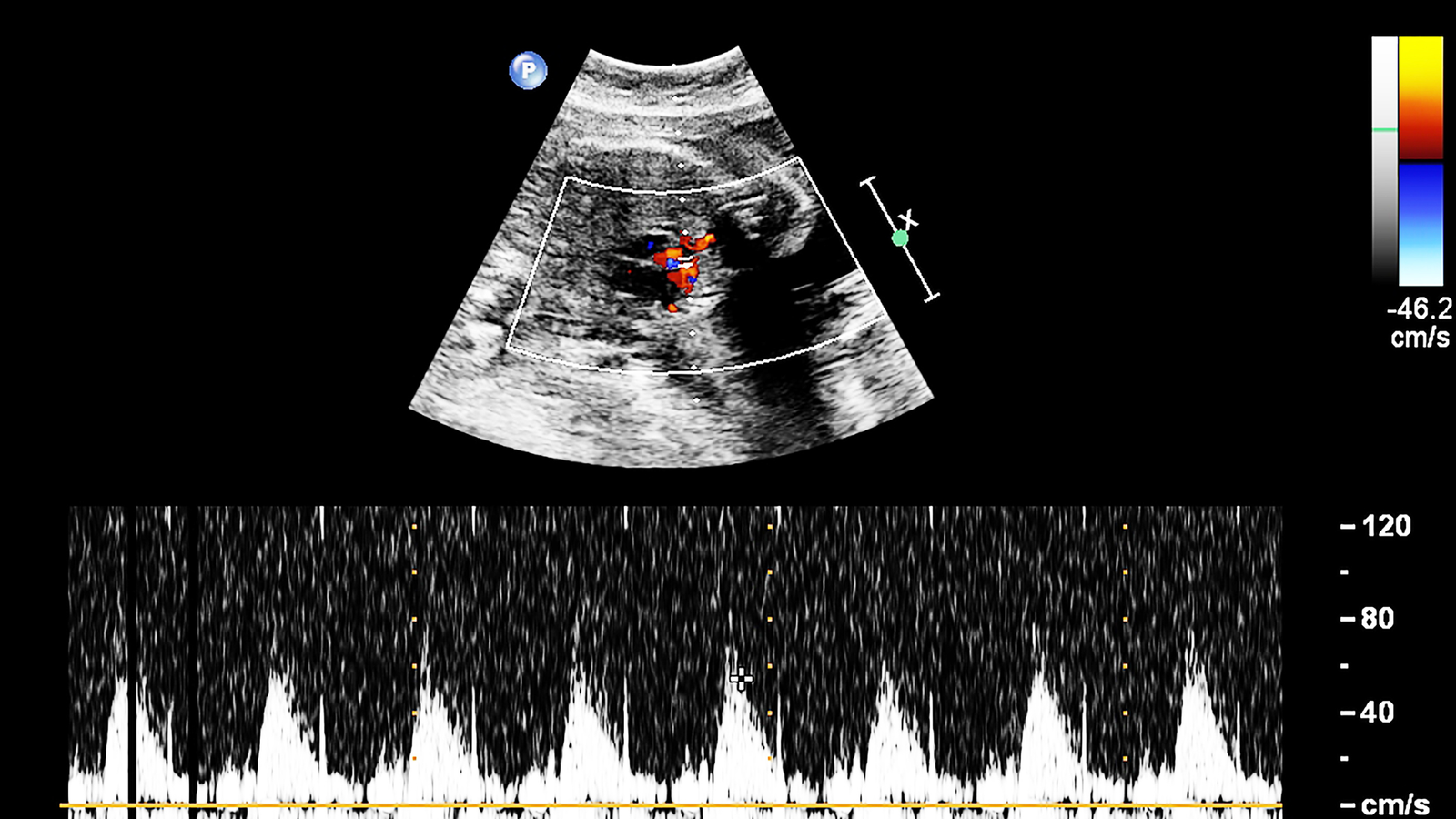
A fetal echocardiogram is an essential diagnostic tool that helps healthcare professionals examine the heart of a developing baby in the womb. This non-invasive ultrasound test offers detailed insights into the fetal heart’s structure, function, and rhythm, allowing early detection of potential heart abnormalities. Early diagnosis and timely intervention are crucial for managing congenital heart conditions and ensuring better outcomes for the baby.
Dr. Nidhi Rawal, one of the best pediatric cardiologists in India with over 15 years of experience, specializes in diagnosing and treating pediatric heart conditions. At Child Heart Health, Dr. Rawal uses advanced fetal echocardiography techniques to provide precise diagnoses and help parents make informed decisions about their baby’s heart health. This blog explores the significance of fetal echocardiograms, what conditions they can diagnose, and how they benefit both the baby and the family.
What is a Fetal Echocardiogram?
A fetal echocardiogram, often referred to as a fetal echo, is a specialized ultrasound designed to assess the structure and function of the fetal heart. It is typically performed during the second trimester, between 18 and 24 weeks of pregnancy, although it can be done earlier or later if necessary.
Unlike a standard prenatal ultrasound that provides a general overview of the baby’s development, a fetal echocardiogram focuses specifically on the heart, offering a more detailed look at the heart’s chambers, valves, blood vessels, and rhythm. The procedure is safe for both the mother and the baby, as it uses sound waves to create images without any harmful radiation.
Why is a Fetal Echocardiogram Performed?
A fetal echocardiogram is recommended if there is a higher risk of congenital heart disease (CHD) in the baby. Some common reasons for conducting a fetal echocardiogram include:
- Family history of congenital heart disease: If either parent or a close relative has a history of heart defects, the risk of the baby having a heart problem increases.
- Abnormal findings on routine ultrasounds: If a routine ultrasound detects potential issues with the fetal heart or other organs, a fetal echocardiogram can provide a more in-depth analysis.
- Maternal health conditions: Certain maternal conditions, such as diabetes, lupus, or infections during pregnancy, may increase the risk of heart problems in the fetus.
- Medication or drug exposure: Some medications or exposure to drugs during pregnancy can increase the chances of heart abnormalities.
- Genetic conditions: If there are concerns about chromosomal abnormalities like Down syndrome, a fetal echocardiogram can help determine if there are any associated heart defects.
What Conditions Can Be Diagnosed with a Fetal Echocardiogram?
Fetal echocardiograms are highly effective in diagnosing a wide range of heart conditions. Early detection allows parents and healthcare providers to plan for any necessary interventions, treatments, or surgeries after birth. Here are some of the most common conditions that can be identified through a fetal echocardiogram:
1. Congenital Heart Disease (CHD)
Congenital heart disease refers to structural defects present in the heart from birth. A fetal echocardiogram can detect various forms of CHD, including:
- Atrial Septal Defect (ASD): A hole in the wall between the two upper chambers (atria) of the heart.
- Ventricular Septal Defect (VSD): A hole in the wall between the two lower chambers (ventricles) of the heart.
- Tetralogy of Fallot (TOF): A combination of four heart defects that affect blood flow.
- Transposition of the Great Arteries (TGA): A condition where the two main arteries leaving the heart are reversed.
- Hypoplastic Left Heart Syndrome (HLHS): A condition in which the left side of the heart is underdeveloped.
- Pulmonary Stenosis: Narrowing of the pulmonary valve, which restricts blood flow from the heart to the lungs.
2. Heart Valve Abnormalities
The fetal echocardiogram can detect abnormalities in the heart valves, such as stenosis (narrowing) or regurgitation (leakage). These conditions can affect how blood flows through the heart and may require intervention after birth.
3. Arrhythmias (Irregular Heartbeat)
A fetal echocardiogram can also detect abnormal heart rhythms, known as arrhythmias. These may include:
- Bradycardia: A slower-than-normal heart rate.
- Tachycardia: A faster-than-normal heart rate.
- Irregular heartbeats: Any abnormal patterns in the heart’s rhythm.
While some arrhythmias resolve on their own, others may require medical attention or monitoring after birth.
4. Cardiomyopathies
Cardiomyopathy refers to diseases of the heart muscle that affect its ability to pump blood effectively. A fetal echocardiogram can help identify conditions like:
- Dilated cardiomyopathy: The heart’s chambers become enlarged, weakening the muscle and reducing its pumping ability.
- Hypertrophic cardiomyopathy: Thickening of the heart muscle, making it harder for the heart to pump blood efficiently.
5. Fetal Heart Tumors
Though rare, fetal echocardiograms can detect tumors in the heart, such as rhabdomyomas, which are non-cancerous growths that can affect heart function. Early detection allows for close monitoring and timely intervention if needed.
How a Fetal Echocardiogram Benefits Your Baby
A fetal echocardiogram provides numerous benefits, ensuring that any potential heart issues are identified early in the pregnancy. This proactive approach allows for:
- Early diagnosis and intervention: Detecting heart abnormalities early enables healthcare providers to plan for medical or surgical interventions immediately after birth, improving the chances of successful treatment.
- Informed decision-making: With a clear diagnosis, parents can make informed decisions regarding their baby’s care and treatment, including potential delivery options and hospital preparations.
- Reduced anxiety: For parents with a family history of heart conditions or other risk factors, a fetal echocardiogram can offer peace of mind or allow for early preparation in case of any detected issues.
What to Expect During a Fetal Echocardiogram
The fetal echocardiogram is a painless, non-invasive procedure similar to a regular ultrasound. Here’s what you can expect during the test:
- Preparation: No special preparation is required for the mother. The procedure is typically performed in a comfortable, dimly lit room to ensure clear images.
- Procedure: A gel will be applied to the mother’s abdomen, and a specialized ultrasound transducer will be used to send sound waves through the body. These waves create detailed images of the fetal heart.
- Duration: The procedure usually takes about 30 minutes to an hour, depending on the position of the baby and the complexity of the heart structures.
- Results: After the test, a pediatric cardiologist like Dr. Nidhi Rawal will review the images and discuss the results with the parents.
Conclusion
A fetal echocardiogram is a powerful diagnostic tool that allows early detection of potential heart problems in the developing baby. For parents, early diagnosis provides valuable information to plan for the best possible care for their child. At Child Heart Health, Dr. Nidhi Rawal, one of India’s top pediatric cardiologists, provides expert care and support for families facing concerns about their baby’s heart health.
If you or your healthcare provider have any concerns about your baby’s heart, schedule a fetal echocardiogram at Child Heart Health today. Early detection is the key to ensuring a healthy start for your little one.


